New interventional cardiology methods revolutionise treatment options. Vascular access is chosen based on clinical circumstances.
‘Interventional cardiology’ refers to diagnostic and therapeutic cardiac procedures performed using a catheter. Interventional cardiology is key to treating cardiovascular disease. It has reduced the mortality rate of patients with conditions such as heart attack, severe heart failure and vascular disease.
Radial artery
Most interventions in coronary arteries are made via catheters (2-3 mm in diameter) inserted through the radial artery (see Figure a). Balloon dilation (PTCA, dilation of a narrowed coronary artery) is the most common procedure in interventional cardiology. It enables rapid and safe treatment of coronary heart disease through implanting a stent (vascular support). Drug-eluting stents (DES) are usually used, as they show a very low restenosis rate. Newer stent technology, such as absorbable stents (BVS) that dissolve over time, is used only selectively. It has yet to prove itself against the established DES in studies and everyday medical practice. Access via the radial artery allows the patient to get up immediately after the procedure and leave hospital on the same day. The risk of bleeding is also lower compared with access via the femoral artery.
Femoral artery
The femoral artery (see Figure b) is the standard access for complex procedures on the coronary arteries. Its larger vascular diameter offers more opportunities for specialised interventions. This access is particularly important in the diagnosis and treatment of heart valve disease. For certain patients, minimally invasive aortic valve replacement (TAVI) is preferable to open heart surgery. Detailed preliminary examinations with ultrasound and computed tomography are required for the success of this procedure. Most TAVI procedures can be performed without general anaesthetic. For the remaining patients, access may occur via the apex of the heart (transapical – see Figure d) or the subclavian artery (see Figure c). Surgery can also be performed on the leg, carotid or renal arteries via the femoral artery.
Femoral vein
Interventions via venous access (see Figure e) involve fewer complications than those via arterial access due to the lower pressure. These interventions include measurement of the blood flow (haemodynamic measurements), tissue samples (cardiac muscle biopsies) and evaluation of pulmonary pressure. Congenital heart defects (shunt defects) are also closed or treated via this access. This intervention risks heart failure, thrombotic complications that may cause a stroke, and a high strain on the circulatory system. Small blood clots in the leg veins may travel into the arterial path and potentially block a cerebral artery. A procedure known as umbrella implantation can prevent these problems – without heart surgery.
Transseptal puncture
Venous access is also used for transseptal puncture (puncturing of the cardiac septum). An opening between the right and left atria is created to allow an intervention in the mitral valve. The MitraClip procedure is a good option for high-risk patients with severe leakage in the mitral valve: it closes the valve with a clip. A successful procedure should combine ultrasound technology with fluoroscopy technology. A hybrid operating theatre offers the optimal conditions for this, and, as with the TAVI procedure, it needs a highly specialised team.
The left atrial appendage (LAA), where blood pools and forms clots in patients with atrial fibrillation, can also be occluded using transseptal puncture. In this technique, the cardiologist closes the left atrial appendage with an implant. The main source of embolism is removed and the risk of stroke reduced, especially for patients who take blood-thinning treatment. After closure, long-term aspirin monotherapy is possible.
Medical progress and healthy lifestyle
These interventional cardiology techniques are a key part of the treatment of cardiovascular disease. They complement progress in heart rhythm management, treatment of heart failure, heart surgery and medication. Thanks to these advances, it is increasingly possible to offer high-quality, minimally invasive treatment to patients. However, patients also require a healthy lifestyle, exercise and a balanced diet for a long, high-quality life.
Article from Prof. Dr. med. Peter Martin Wenaweser, specialist in cardiology and general internal medicina.
GLOSSARY
• Minimally invasive: intervention via a small cut in the skin instead of open surgery
• Cardiovascular: affecting the heart and vascular system
• Ballon dilation: widening of a narrow artery using a balloon catheter
• Stent: vascular support
• Tavi: minimally invasive aortic valve replacement (Transcatheter Aortic Valve Implantation)
• Transseptal puncture: puncturing of the cardiac septum
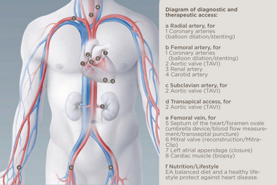
Diagram of diagnostic and therapeutic access:
a Radial artery, for
1 Coronary arteries
(balloon dilation_/_stenting)
b Femoral artery, for
1 Coronary arteries
(balloon dilation_/_stenting)
2 Aortic valve (TAVI)
3 Renal artery
4 Carotid artery
c Subclavian artery, for
2 Aortic valve (TAVI)
d Transapical access, for
2 Aortic valve (TAVI)
e Femoral vein, for
5 Septum of the heart_/_foramen ovale
(umbrella device/blood flow measurement/transseptal puncture)
6 Mitral valve (reconstruction_/_MitraClip)
7 Left atrial appendage (closure)
8 Cardiac muscle (biopsy)
f Nutrition_/_Lifestyle
A balanced diet and a healthy lifestyle protect against heart disease.